The Fusiform Face Area: Why Your Brain Has a Dedicated Face-Perception Module
In 1997, Nancy Kanwisher's lab identified a small region of the fusiform gyrus — the FFA — that responds more strongly to faces than virtually anything else. This article unpacks what the FFA is, where it sits, what it actually computes, and why a 2020 finding (blind people have an FFA too) is still rattling assumptions about how the brain organizes itself.

In 1997, Nancy Kanwisher and her colleagues published a nine-page paper in the Journal of Neuroscience that would become one of the most cited findings in cognitive neuroscience: the identification of the fusiform face area (FFA), a region in the fusiform gyrus that responds more strongly to faces than to virtually anything else the brain encounters.1 The paper has since accumulated over 10,000 citations, but its real legacy is the question it planted: does the brain contain specialized, dedicated modules for specific categories of thought?
Where it is and what it does
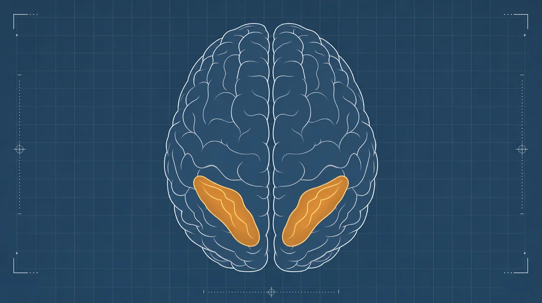
The FFA sits on the lateral surface of the mid-fusiform gyrus, a fold of cortex running along the bottom (ventral) surface of the temporal lobe. In most right-handed people it is more pronounced in the right hemisphere, though a left-hemisphere version shows up in roughly half of individuals.
The defining property is its preferential response to faces over objects. In the original 1997 study, 12 of 15 subjects showed a region in the right fusiform gyrus where the fMRI BOLD signal was significantly higher during viewing of face photographs than of assorted common objects. The region was small — about 1 cubic centimeter on average, located at roughly Talairach coordinates 40, −55, −10 in the right hemisphere — yet its selectivity was striking.1
What made the finding rigorous was the multi-test approach. Kanwisher's team didn't stop at one faces-vs.-objects comparison. Using the same functionally defined region of interest in each subject, they ran four additional tests designed to rule out alternative explanations:
- Intact vs. scrambled two-tone faces — rules out low-level luminance or edge cues
- Faces vs. houses — shows the region responds to faces specifically, not to "sets of similar exemplars"
- 3/4-view faces (hair concealed) vs. hands — rules out hair cues or general response to human body parts
- 1-back matching task on faces vs. hands — rules out visual attention as the driver (the hand task was equally or more difficult)
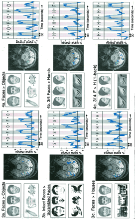
All four comparisons showed the same result: higher FFA response for faces. The systematic elimination of alternative hypotheses through repeated testing on the same functionally defined region was itself a methodological innovation — the functional region of interest (fROI) approach — that reshaped how cognitive neuroscience experiments are designed.2
What the FFA actually represents
By the time of the 2006 review by Kanwisher and Yovel, substantial evidence had accumulated about what the FFA is actually computing.3
It responds to faces, not to simple visual features. The FFA lights up for front-view photos, profile views, line drawings, cat faces, and two-tone "Mooney faces" — stimuli that share almost no low-level features. If you take a bistable image (like the classic vase/face illusion) and track what subjects perceive moment-by-moment, the FFA response tracks conscious face perception even though the retinal input is unchanged.
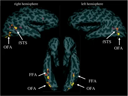
It is implicated in face identity, not expression or gaze. Functional dissociations show the FFA responds strongly during tasks requiring discrimination of who a face belongs to. A region in the superior temporal sulcus (fSTS) handles changeable face properties like gaze direction and emotional expression. Damage to fusiform cortex — the acquired syndrome of prosopagnosia — selectively destroys the ability to recognize faces while leaving object recognition largely intact, a double dissociation that points to a domain-specific machinery.
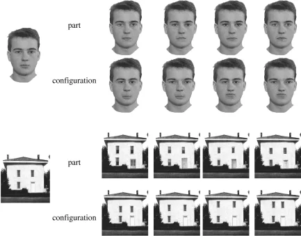
It processes faces holistically. The FFA shows a "face inversion effect" in neural terms: a higher BOLD response for upright than inverted faces, and this neural inversion effect correlates across subjects with the behavioral face inversion effect. It also shows fMRI evidence of the "composite face effect" — the tendency to process two halves of a face as a unified whole rather than independently.
The main controversy: is the FFA really face-specific?
Two challenges have occupied the field for over two decades.
The expertise hypothesis (Gauthier, Tarr and colleagues) proposed that the FFA is not truly face-specific, but rather supports fine-grained discrimination of any category for which one has acquired substantial visual expertise — cars for car experts, birds for birders. The idea draws intuitive appeal from the fact that we are all, in a sense, "face experts." However, repeated attempts to produce FFA expertise effects with novel stimuli have mostly failed or yielded small effects that are not restricted to the FFA. The most systematic tests — training subjects for many hours on novel objects that look nothing like faces — find no significant FFA increase for trained over untrained objects, but do find training effects in a more general object-selective region (the lateral occipital complex).3
The distributed representation challenge (Haxby et al., 2001) made a different point: the pattern of response across voxels in the FFA — not just the mean response — contains information about non-face object categories. Multi-Voxel Pattern Analysis (MVPA) can decode whether you're looking at a chair or a shoe from FFA activity patterns, even though the region responds weakly to both. Kanwisher's position is that what matters is not the information scientists can extract from the region, but what information the rest of the brain reads out from it. Lesion and stimulation studies — which test actual causal necessity — show the FFA is causally needed for face processing but not for objects. A 2012 study using direct electrical stimulation of fusiform face-selective regions demonstrated that stimulation distorts face perception, confirming the causal role.2
One finding that surprised everyone: blind people have an FFA too
In 2020, MIT researchers reported that people who have been blind since birth — and have therefore never seen a face — still show face-selective activation in the fusiform gyrus.4 The FFA activates when blind individuals touch a face, and also in response to information received through other senses. This result challenges any straightforward experiential account of the FFA and suggests the region's location and tuning may be at least partly innately constrained — not learned, but ready to be recruited by whatever sensory inputs deliver socially relevant identity information.
Why it matters beyond face perception
The FFA's significance is not only that it answers questions about face recognition. It is that it demonstrated — in a replicable, methodologically rigorous way — that discrete regions of the human cortex can be dedicated to surprisingly specific cognitive functions. The localize-and-test fROI method developed in the FFA paper became the standard approach for discovering other category-selective regions: the parahippocampal place area (PPA) for scenes, the extrastriate body area (EBA) for bodies, the visual word form area for written language, and the temporoparietal junction (TPJ) for thinking about other minds.
The broader conceptual bet the FFA placed — that the human brain is not a general-purpose processor but a collection of specialized modules — remains an active question. But the fusiform face area gave that debate its clearest concrete example, and subsequent research in macaques (where single-unit recordings confirmed that 97% of neurons in fMRI-identified face patches respond exclusively to faces) has strengthened the case that the selectivity is real, sharp, and evolutionarily old.3
Landmark paper: Kanwisher, N., McDermott, J. & Chun, M.M. (1997). The fusiform face area: a module in human extrastriate cortex specialized for face perception. Journal of Neuroscience, 17(11), 4302–4311. Read at JNeurosci
Course connection: MIT 9.13 (The Human Brain) uses the FFA as the central case study for functional specificity and the modular organization of the brain. Kanwisher's 2010 PNAS paper "Functional specificity in the human brain" is the Week 1 required reading for the course.5
References
- 1Kanwisher et al., 1997 — The Fusiform Face Area
- 2Kanwisher, 2017 — The Quest for the FFA and Where It Led
- 3Kanwisher & Yovel, 2006 — The Fusiform Face Area: a cortical region specialized for face perception
- 4MIT News, 2020 — Face-specific brain area responds to faces even in people blind from birth
- 5MIT OCW 9.13 Readings
Add more perspectives or context around this Post.